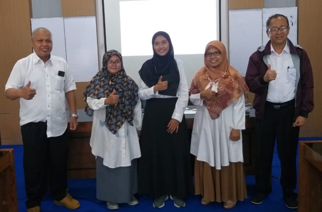
SIDANG SKRIPSI DAN SIDANG TESIS JANUARI 2023
Bagi tiga anggota Plasma RG, bulan ini merupakan bulan bahagia untuk mereka karena telah melaksanakan Sidang Skripsi dan Sidang Tesis....
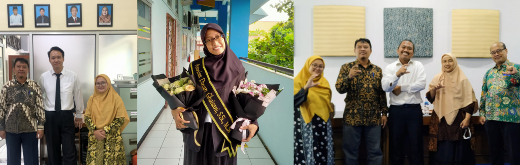
Bagi tiga anggota Plasma RG, bulan ini merupakan bulan bahagia untuk mereka karena telah melaksanakan Sidang Skripsi dan Sidang Tesis....
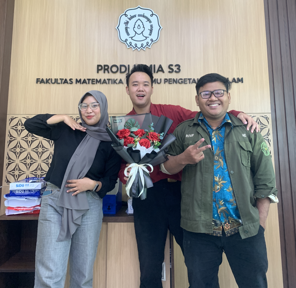
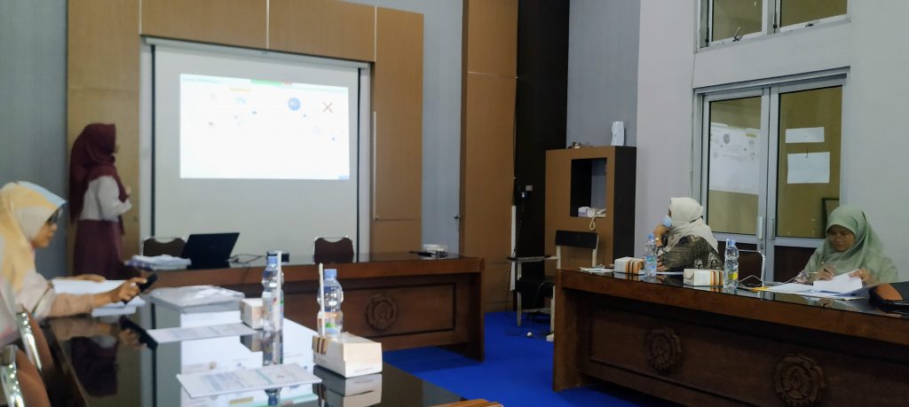
Bulan Januari ini, dua anggota dari Plasma RG yaitu Umam Hasan Setiawan dan Yopi Santoso melaksanakan Seminar Hasil Skripsi dan...
Annisa Dinan Ghaisani merupakan mahasiswa Magister Kimia UNS yang menjadi anggota Plasma RG. Pada 23 Desember 2022 yang lalu, Dinan telah mempresentasikan hasil...
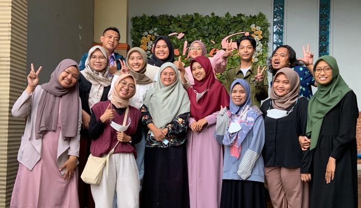
Ada yang pergi ada yang datang, begitulah hidup. Pada tanggal 21 Desember 2022 yang lalu, Plasma RG mengadakan Farewell &...
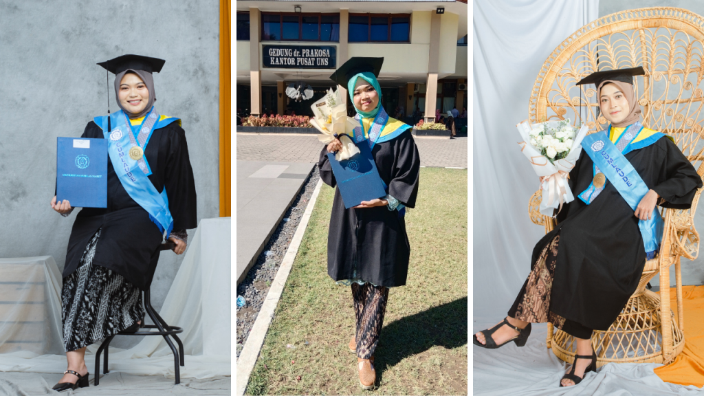
Di penghujung tahun ini banyak kabar gembira yang menghampiri Plasma RG, salah satunya adalah wisuda dari beberapa anggota Plasma RG....
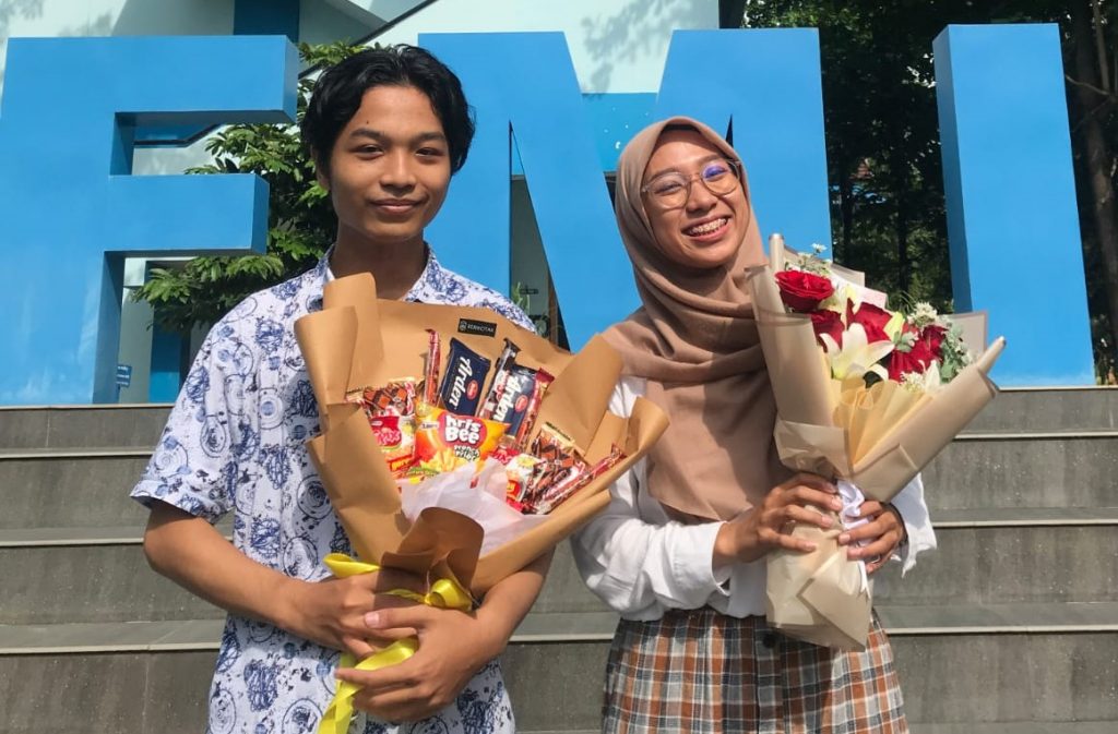
November menjadi bulan yang penting bagi dua anggota baru Plasma RG yang merupakan mahasiswa Prodi Kimia angkatan tahun 2019 yaitu...
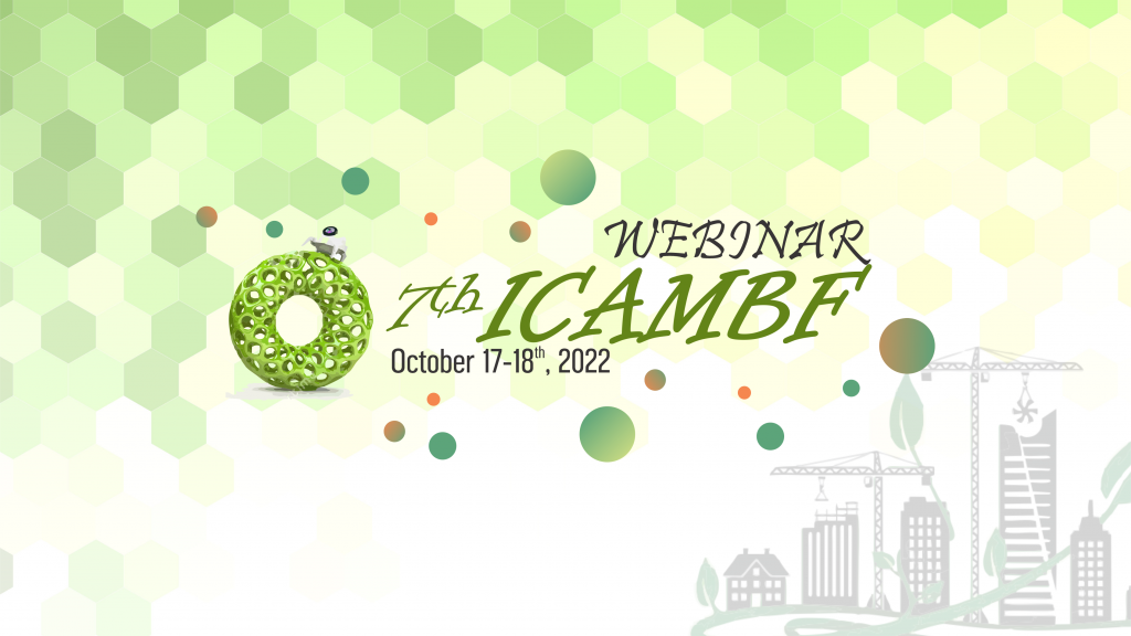
Pada tanggal 17 & 18 Oktober 2022, beberapa anggota Plasma RG mempresentasikan hasil penelitian mereka pada Acara Webinar 7th International...
Salah satu anggota Plasma RG telah melakukan sidang skripsinya pada tanggal 17 Oktober 2022. Dia adalah Megadita Ayuningtyas, mahasiswa Fisika...
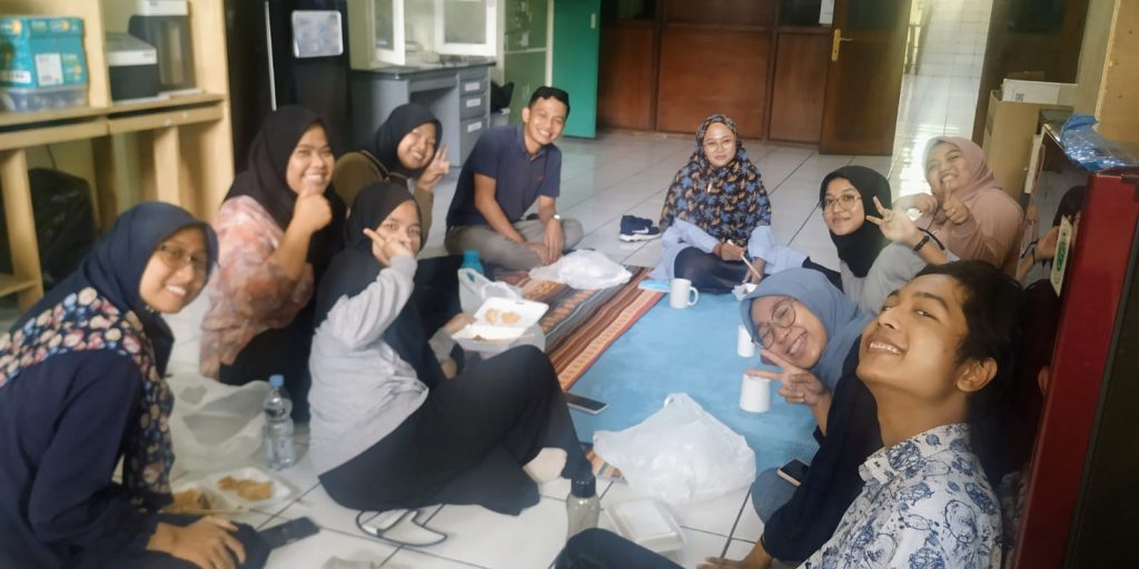
Pada tanggal 11 Oktober 2022 merupakan hari yang menyenangkan bagi anggota Plasma RG. Pada siang itu mereka mengadakan makan siang...
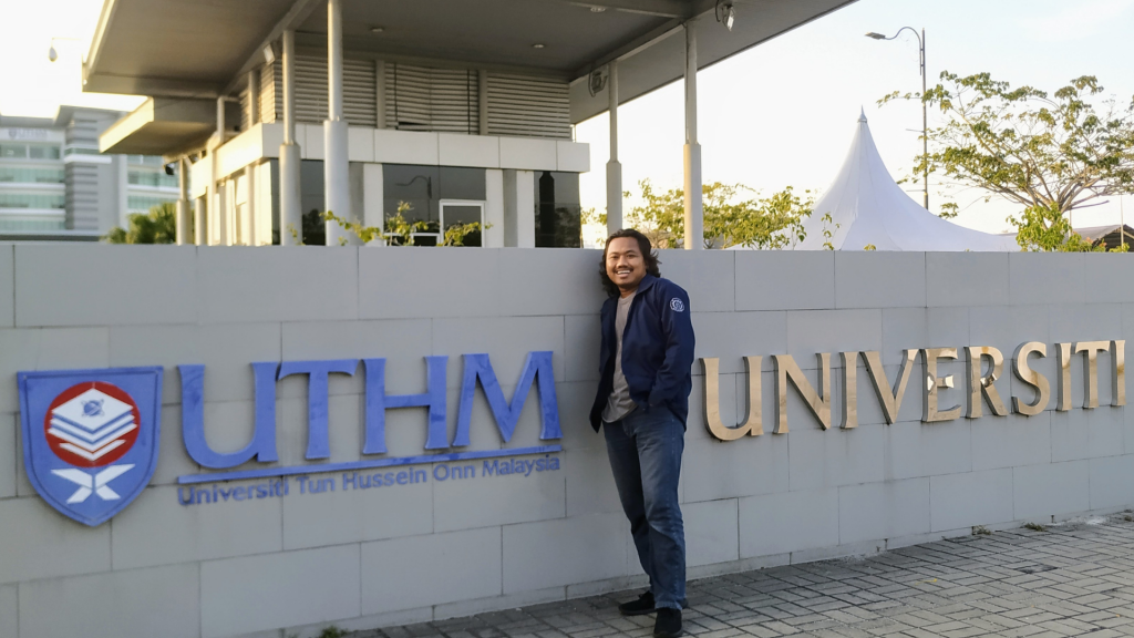
Melalui Global Challenge 2022 yang diadakan oleh UNS, salah satu anggota Plasma RG mendapat kesempatan untuk melakukan visiting research di...
![]() Plasma Science & Technology RG
Plasma Science & Technology RG
Department of Chemistry, Faculty of Mathematics and Natural Sciences, Sebelas Maret University
Jalan Ir. Sutami No. 36A, Kota Surakarta, Jawa Tengah 57126, Indonesia
Locate us on GMAPS
Copyright © 2022 | teguh.staff.uns.ac.id